CHAPTER 1 • LESSON 4 • INVESTIGATE
Why do some bacteria cause us problems?
PURPOSE | To figure out science ideas that help us toward answering our Driving Question by gathering and making sense of evidence.

TIME
Two 50-minute class periods
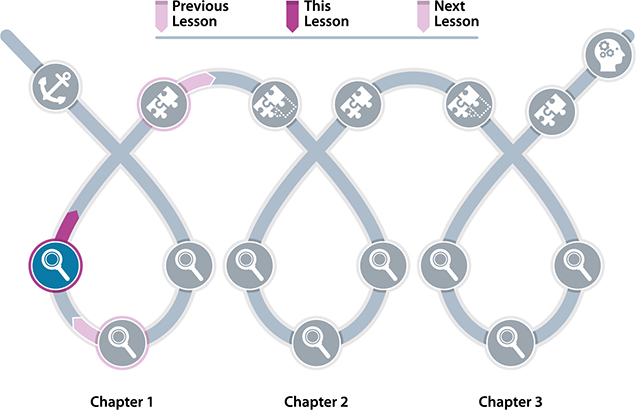
PREVIOUS LESSON | We investigated what bacteria need to live and how they grow. This left us wondering why bacterial growth can sometimes cause us problems.
THIS LESSON | We figure out that bacteria are on us and in us all the time, but they only cause symptoms when particular types grow and reproduce in particular environments. This leaves us wondering how this process worked in Zach’s case and whether it always works the same way to cause symptoms.
NEXT LESSON | We will develop a model to figure out how all the ideas from this chapter connect with each other to explain how and why bacteria can sometimes make us sick. This will leave us wondering: How can we get better?
 LESSON LEARNING GOAL
Compare, integrate, and summarize sources of information to explain how bacteria growing and reproducing in environments within humans can cause us to experience symptoms.
Lesson question
Why do some bacteria cause us problems?
What students figure out
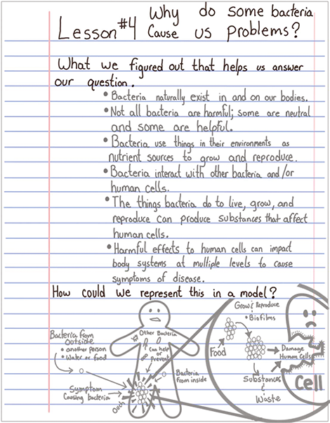
Bacteria naturally exist in and on our bodies.
Not all bacteria are harmful; some are neutral and some are helpful.
Bacteria use things in their environments as nutrient sources to grow and reproduce.
Bacteria interact with other bacteria and/or human cells.
The things bacteria do to live, grow, and reproduce, and the substances they produce can affect human cells.
Harmful effects to human cells can impact body systems at multiple levels to cause symptoms of disease.
What we are not expecting
Where we are not going yet
We are not yet investigating how the human immune system responds to bacteria.
Boundaries
We do not expect students to memorize which types of bacteria cause various diseases and how.
Relevant common student ideas
Only certain tissues, like blood or brain tissue, are made of cells. (All tissues are made of cells.)
Animal cells are typically round. (The shape and structure of cells [from any organism] vary depending on their functions. Specialized cells may have unique shapes.)
Key literacy and sensemaking strategies
-
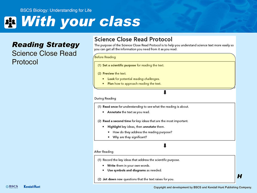
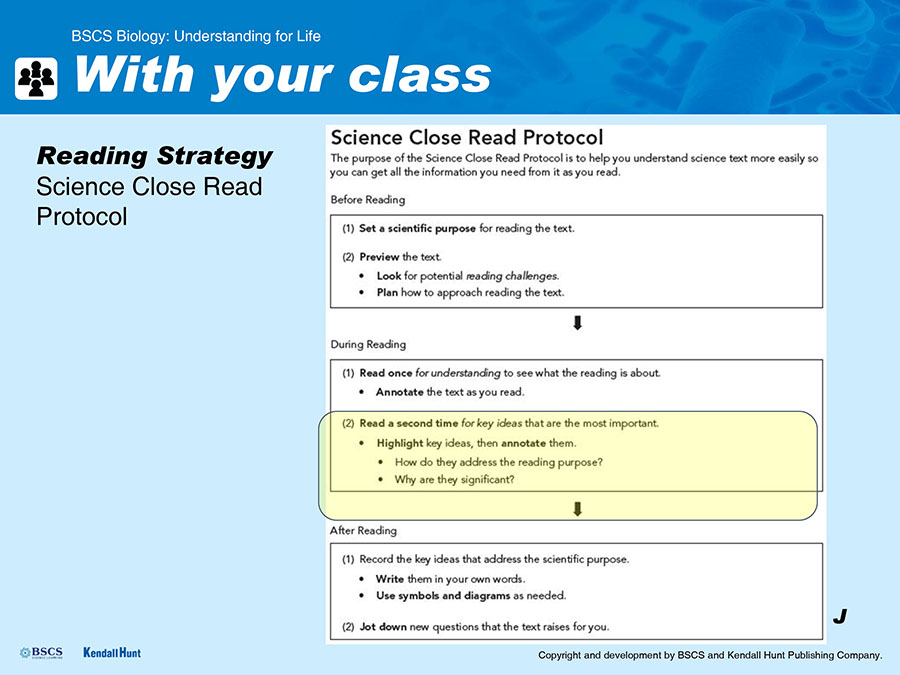
Students use the Science Close Read Protocol for the first time in the unit. The entire protocol will be modeled for them first, then used by them in small groups.
Student Science Reading Annotation Stems
Students have used the science reading annotation stems previously in Lesson 3. They will leverage their use in the close read protocol.
-
Giving and Receiving Feedback is used for a second time as students review each other’s charts and diagrams. Students will review and use the strategies that were introduced in Lesson 2, with an emphasis on receiving feedback.
Model Tracker
The Model Tracker is the primary strategy students use to clarify and capture their individual sensemaking in this lesson.
Model Tracker Self-Assessment and Feedback Tool
The Model Tracker Self-Assessment and Feedback Tool is used as it was previously in Lessons 2 and 3.
Model Tracker Formative Assessment Tool
The Model Tracker Formative Assessment Tool is used by the teacher for the first time as a way to assess students’ progress in content knowledge and in using modeling practices.
ASSESSMENT
Formative check: Model Tracker
After completing this lesson and before the next (Synthesize) lesson, use the Model Tracker Formative Assessment tool to assess individual student progress across this chapter, and whole-class readiness for the Synthesize Lesson.
Examine the Model Tracker entries for Lessons 2, 3, and 4 in student notebooks. The Model Tracker Formative Assessment Tool provides guidance on what to look for in student Model Trackers across all three of the lessons in the form of a checklist. As you work through the checklist keep in mind that you are looking for:
The presence or absence of components (not the quality of the representations)
Interactions that may be represented either in verbal descriptions or with diagrammatic elements such as arrows or lines
Interactions shown as arrows or other directional indicators showing inputs and outputs of bacterial cells (or other subsystems)
Interactions shown as arrows or otherwise indicating cause-and-effect relationships
Boundaries of subsystems in models from Lesson 4. These may not be explicitly indicated or labeled as system boundaries, but may be indicated simply by separating one subsystem (bacterial cell, human cell, tissue or organ that is affected) from another graphically or spatially. Subsystems should have indications of interactions between them.
Use of the model to explain something. Each lesson’s model will explain something a little bit different. The models should show some attempt to answer the lesson questions. For instance, a labeled bacterial cell alone is not sufficient in Lesson 2, rather the model should show some indication of how bacteria might grow.
Also keep in mind that you are NOT looking for neatness or for accuracy in the Model Tracker. The models students draw here are a description of their growing conceptual understanding and as such they may be inaccurate in some details of components, interactions, or explanations of phenomena. In the appropriate space in each student’s Model Tracker Self-Assessment and Feedback Tool in the front of their science notebook, provide feedback to students regarding the models in their Model Tracker for Lessons 2–4. Give students feedback on:
Any missing, or extraneous components
The absence of any interactions that are described in the Assessment Tool
The directional nature of cause-and-effect relationships
Whether or not they have used the model to attempt to explain a phenomenon or answer a question (instead of simply labeling a diagram)
Any particularly effective elements of their models that helped you understand their thinking (Whether or not their thinking is accurate, the way they organize their models is as important as the accuracy of what they represent. This is why the feedback section has separate spaces for feedback on content and modeling.) This feedback will help students improve their practice of modeling and support continued growth in their conceptual understanding.
 INVESTIGATE LESSON SNAPSHOT
Lesson 4: Why do some bacteria cause us problems?
BIG IDEA | Some (but not all) bacteria can cause us problems when they enter our bodies (or a new place in our bodies) because in the process of getting what they need to live and grow, they may damage our cells and cause us symptoms of infection.
Routine |
Part |
Time |
Summary |
Slide |
Materials |
|
1 |
10 min |
Consider why bacteria growth might cause problems Students reflect on what they have learned in Lessons 1 and 2 and consider how bacteria grow in and on humans and why this might sometimes cause us problems. Purpose: to co-construct the lesson question. |
A–C |
Science notebook |
|
2 |
5 min |
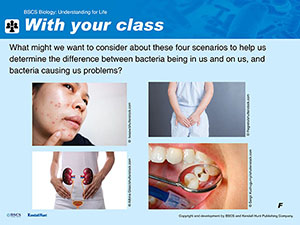
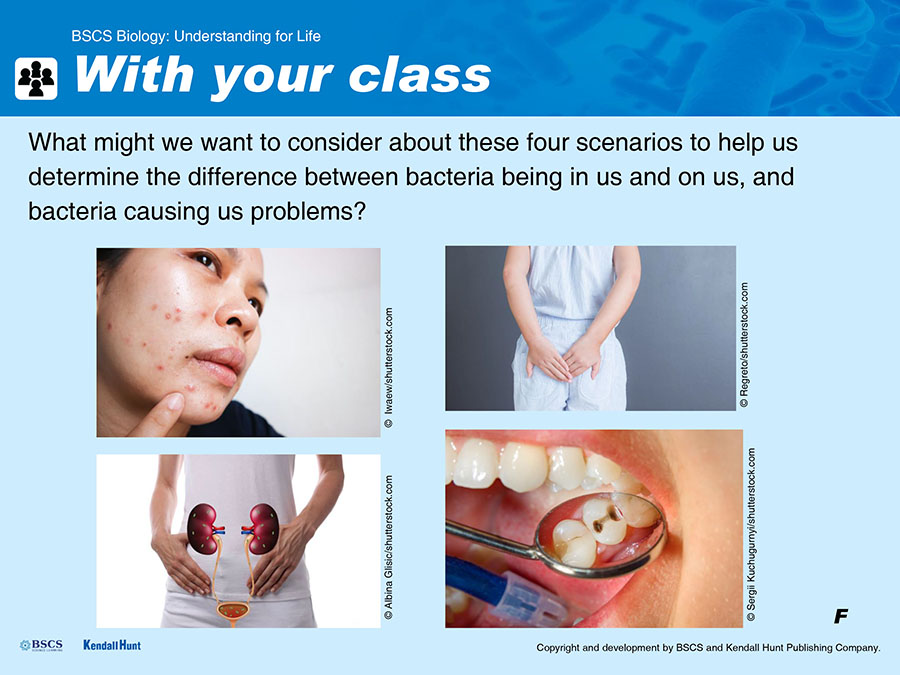
Orient to four different case scenarios and asking questions Students participate in a discussion regarding four different case scenarios (bacteria in the gut, bacteria in the mouth, bacteria in the urinary tract, bacteria on the skin) that they will read to determine the difference between bacteria being in and on humans and an infection that causes symptoms. Purpose: to identify what information to look for in the readings. |
D–F |
|
3 |
20 min |
Model the use of the Science Close Read Protocol Students follow along as you model the use of the close-reading protocol with the first scenario (bacteria in the gut). After you model each element of the close-reading protocol, students have the opportunity to try the strategies and share their ideas as a class. Purpose: to introduce and model the Science Close Read Protocol and have students practice the protocol using one of the readings and a graphic organizer. |
G–L |
Science notebook Student Sheet 1.4.F: Science Close Read Protocol Student Sheet 1.4.A: Gut Feelings Student Sheet 1.2.E: Science Annotation Reading Stems (in the science notebooks) |
|
4 |
15 min |
Small-groups of students use the Science Close Read Protocol Students work in small groups (three to four students) to read one of the three other scenarios (bacteria in the mouth, bacteria in the urinary tract, bacteria on the skin) using the close-reading protocol and a graphic organizer. Purpose: for students to gather information and identify key ideas that will allow them to consider the difference between bacteria being in us and on us and an infection that causes symptoms. |
M–N |
Student Sheet 1.4.B: Bacteria in the Mouth Student Sheet 1.4.C: Myth Busted Student Sheet 1.4.D: What Really Causes Acne? Science notebook |
|
Suggested class period break End of Day 1 |
|||||
|
|
5 |
15 min |
Summarize findings across cases. Students review briefly then reorganize into jigsaw groups of three to summarize the findings for all the cases on chart paper. Purpose: to summarize, paraphrase, and communicate the key ideas from their cases. |
O–P |
Completed Student Sheet 1.4.E: Recording Key Ideas (from day 1) |
6 |
17 min |
Generate representations of general key ideas. In their jigsaw groups, students create diagrammatic representations of the key ideas as they apply to all the cases, then provide peer feedback to other groups. Purpose: to have students engage with modeling as a support for sensemaking, and to lay the groundwork for the Synthesize Lesson that follows |
Q–T |
Science notebook Sticky notes Fine-tip markers |
|
7 |
10 min |
Update Model Trackers. Students fill in their Model Trackers individually. Purpose: to allow students to create summary models independently and keep track of what they figured out. |
U–V |
Science notebook |
|
|
8 |
5 min |
Reflect on what caused Zach’s illness. Students reflect on how their models might connect to the timeline of Zach’s illness. Purpose: to have students identify that they have some ideas about what caused Zach’s illness, but they still have many questions. |
||
9 |
3 min |
Record new questions. Students reflect on and record any new questions they may have about Zach’s case as a result of what they learned in Lesson 4 and add them to the Driving Question Board (DQB). Purpose: to refocus students’ attention on questions as they prepare for the Synthesize Lesson that follows. |
Sticky notes Driving Question Board |
||


LESSON MATERIALS
Per student
Science Notebook
Sticky notes
Fine-point markers
For jigsaw*
8 copies of Student Sheet 1.4.B: Bacteria in the Mouth
8 copies of Student Sheet 1.4.C: Myth Busted
8 copies of Student Sheet 1.4.D: What Really Causes Acne
*Assuming a class size of 32.
Per class
Computer with projector
Markers
Per group of 4 students
Chart paper
Preparation
Clear a space in your classroom for students to display chart papers.
Group students into groups of 3–4 to close read individual scenarios.
Consider how you will create jigsaw groups composed of 4 students (in which each student has read a different one of the four scenarios).
Make copies of student sheets.
ADDITIONAL CONTENT BACKGROUND FOR THE TEACHER
Bacteria, viruses, and fungi are everywhere in the world—on virtually every surface and in virtually all the air we breathe. Our bodies are covered by bacteria, both inside and out. However, the vast majority of these are harmless, or even beneficial to people (and animals), and never cause disease or symptoms of any kind. These bacteria live, grow, and reproduce on and in our bodies all the time without our being aware of them. Students may need to be reassured that these frequent interactions with harmless bacteria are a normal part of living on Earth, and there is no reason to be concerned about them.
A small subset of bacteria (or viruses, or fungi) are pathogenic. That is, they can cause disease under some circumstances. When pathogenic bacteria enter into an environment on or in the body where they have the food and space they need to grow and reproduce sufficiently, they cause symptoms of disease. This is called an infection. These symptoms arise from the specific interactions of the pathogenic bacterial cells and their waste and other products with the body cells, tissues, organs, and systems. The damage caused by the bacteria, and the body’s reaction to that damage, result in the symptoms that people experience.
Bacterial species such as E. coli, P. acnes, or S. mutans often have several different “strains” that are distinguishable biochemically. In a single species, there may be some strains that are pathogenic and some that are not. So, for instance, only certain strains of E. coli can cause disease symptoms in people. Other E. coli strains can grow on us and in us normally without causing any symptoms.
Bacteria that cause infections in people can usually be passed from person to person either directly (e.g., by touch or by sharing of bodily fluids) or indirectly (e.g., by fecal material contaminating a water source that is used to wash food that is consumed without being cooked). Occasionally, bacteria that are pathogenic to humans can grow in an environment outside the body, and may be passed from that environment to a person where they cause an infection.
 NAVIGATE
PURPOSE | To maintain coherence and continuity
Students reflect on our progress so far.
Display Slide A. Have students open their science notebooks to a new page and write the date and “Lesson 4.” Remind them that we’ll add the lesson question once we’ve decided on it. Ask students to think about what we learned in Lessons 2 and 3 and quietly consider the prompt on Slide A.
Students discuss bacterial growth in and on humans.
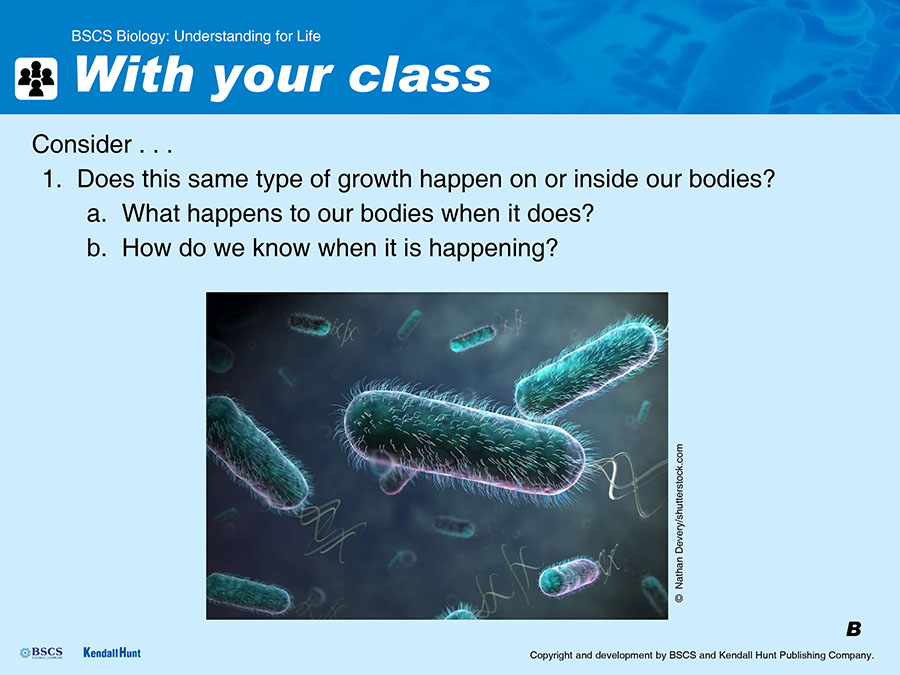
Display Slide B. Once students have described a few concepts they took away from Lessons 2 and 3, ask students to think about what bacterial growth on or inside a human environment might look like. Accept all answers, however, it is important that at least some of the ideas they share include that bacteria do not always cause problems, but can be neutral or even helpful. If necessary, probe to draw out those ideas.

| Suggested prompts | Listen for student responses such as |
|---|---|
| Does this same type of growth happen on or inside our bodies? |
|
| What happens to our bodies when bacteria grow on us or in us? |
|
|
Do we know when it’s happening? How do we know? |
|
Draw attention to the idea of symptoms.
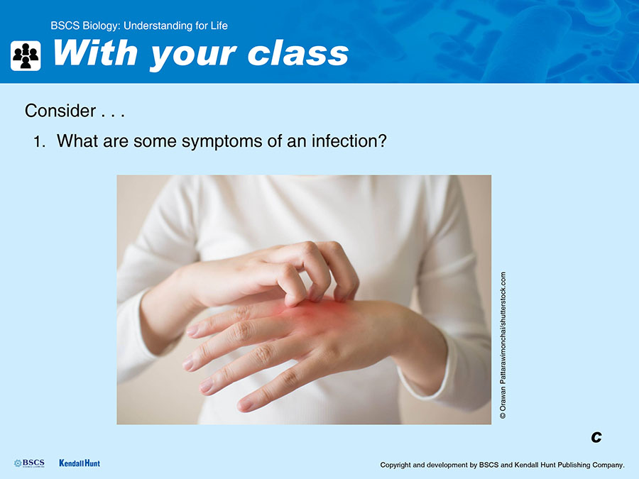
Display Slide C. Lead a whole-class discussion to surface and elicit their ideas about what symptoms (problems, or changes in feeling or function) they’d feel if they were “infected” (e.g., pain, itchiness, redness). The idea of symptoms will have come up in Lesson 1 when students first learned about Zach’s MRSA story. At this point, work with students to add “symptoms” to the Word Wall, and leverage students’ own experiences as well as their observations from Zach’s case to include examples.

Students co-construct the lesson question.
Ask students what they think will be important to figure out in this lesson in order to investigate how bacteria interact with our bodies. Using their suggestions, capture a question in their language that is similar to Why do some bacteria cause us problems? and post it on a whiteboard or other publicly visible space. Have students record the question on the page they started for Lesson 4 in their science notebooks.
 GATHER EVIDENCE
PURPOSE | To plan and carry out evidence gathering tasks
Students orient to examining scenarios.
Ask students to consider what information might be helpful in determining the difference between bacteria being in us and on us and when they cause us problems.
Suggested framing: “So we have some uncertainty in terms of when we think some bacteria can cause us problems, and why. What information might we need to help us figure this out?”
Students are likely to suggest looking at some kinds of infections or infectious diseases that are caused by bacteria.
Note: Students will likely use the word “infection” or refer to “infectious diseases.” At this point these are words they encounter. They can be added to the words we encounter list. These are words they will earn by the end of the lesson, after they distinguish between nonpathogenic bacterial growth (bacteria in us and on us but not causing harm) and pathogenic bacterial growth that causes symptoms or problems (infection or infectious diseases).
Inventory our personal experiences with the scenarios listed.
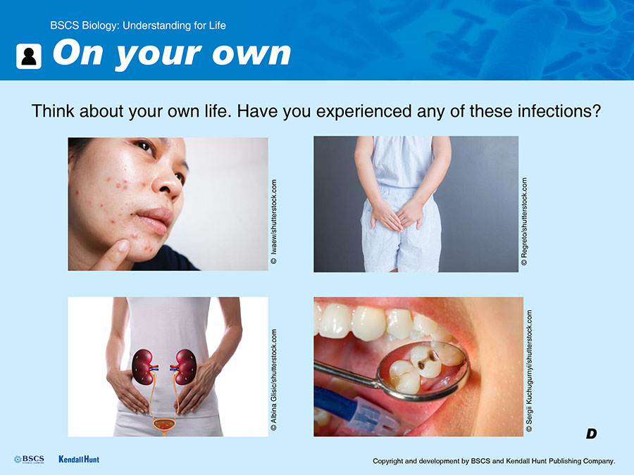
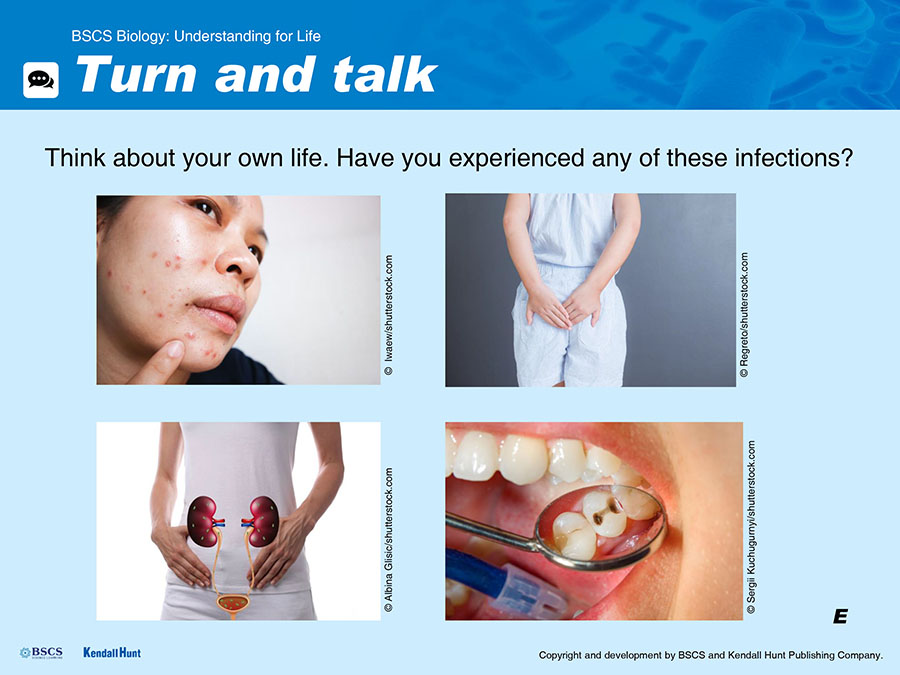
Display Slide D. Ask students to look at the scenarios listed on Slide D and think about whether they have any personal experience (their own, or a family member’s or friend’s) with any of them. Let students know they will have some time to think individually and then they will share with a partner. Knowing in advance that they will be talking about the experience will help students choose an experience they are comfortable sharing with others. Once students have had some time to think on their own, display Slide E and invite students to turn and talk with a partner.
Attending to Equity
Students may differ in their comfort sharing personal experiences with infections. Make sure they know in advance that they will be sharing with partners, and that it is clear to students that they do not need to share any experience if they are not comfortable doing so.
Students think about how to use the scenarios to answer the lesson question.
Display Slide F. Facilitate a discussion using questioning to elicit students’ ideas about what details might be helpful in determining the difference between having bacteria in us and on us vs. having bacteria when they cause us problems.
Suggested prompts:
What might we want to look for in these scenarios?
Are the bacteria just doing what they naturally do to survive when they are in us or on us or are they doing something else?
If they are just surviving, what do the bacteria need to survive?
Summarize student ideas in the form of questions they want to answer when considering the scenarios:
What kind of bacteria are there in this environment in the body?
Do all the bacteria in the environment cause symptoms (an infection)?
How many bacteria are there?
How did they get there?
What is the bacteria’s food source and what waste products or other substances do they produce?
How do they interact with other cells?
Capture these questions on a whiteboard or other publicly visible space. Have students record the questions on the page they started in their science notebooks.
Attending to Student Ideas
This discussion allows you to provide a support opportunity for students to realize there are gaps in their understanding of what it means to have symptoms or have an infection and to surface the types of questions they want to try to answer. During this discussion, encourage all students to share their ideas and experiences and clarify their ideas.
Developing the Practices: Asking Questions
During high school, students should make progress on an ability to ask questions that can be answered within the scope of available resources. Push them to propose questions that could be answered using information they might obtain by reading a text containing scientific information at a high school level.
Explain to students that we will use the jigsaw strategy to work on the scenarios and then share our findings with others, but that first we will model reading a text with the Science Close Read Protocol.
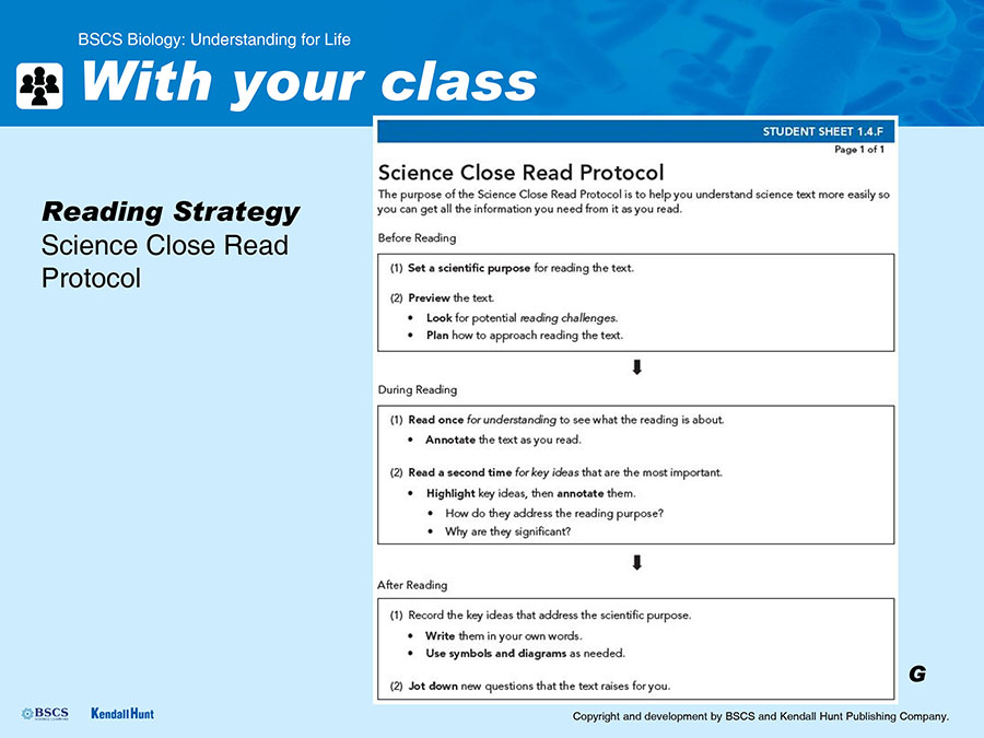
Display Slide G. If necessary, explain to students that a jigsaw strategy entails each individual working with a small group to become an expert on one of the scenarios, then joining a different group in which each student is an expert on a different scenario to share and communicate ideas and information with others. Distribute Student Sheet 1.4.F: Science Close Read Protocol, and have students put it in their science notebooks (in the back with their other course-level resources) so they can refer to it during this lesson and in the future.

Provide students with a rationale for using the Science Close Read Protocol.
The text describing the scenarios will likely be challenging for some students, and the Science Close Read Protocol is designed to support students to become more comfortable and successful when reading scientific texts. Providing an explicit rationale as to why students should use the strategy will help students understand its purpose. You might say something like, “Reading is important in science because much scientific information is communicated in texts. There are many different kinds of science texts because there is so much great information in science. The Science Close Read Protocol helps us to understand the information so we can act on it.”
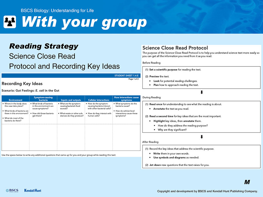
Briefly introduce the Science Close Read Protocol.
Display Slide G. Point out the three major sections (Before, During, and After Reading) and the steps within each section. Tell students you will model the Science Close Read Protocol for them as they follow along, and then they will be using it themselves. Say something like, “A critical part of ‘doing science’ is trying to figure out what I am reading and why I am reading it. Today, I’m going to show you how I do it and what’s helpful for me.”
Literacy and Multilingual Learner Support
Modeling reading strategies: modeling your own reading processes is a helpful way to introduce your students to the literacy tasks involved in science readings. The best opportunities to model strategies for your students include:
When the text requires students to use a strategy they are not already using.
When the text has a new or especially tricky literacy challenge.
When you are using the science close read protocol, especially the first time you use it.
Although there are many reading strategies for your students to learn, it is best for you to model a single strategy at a time when it is particularly important for students to use it with the text they are reading (e.g., making thinking visible with annotations, setting a science reading purpose). This modeling demonstration can be brief but purposeful.
Supporting students to use the Science Close Read Protocol: The purpose of the Science Close Read Protocol is to support reading comprehension, which includes understanding the text and applying the information.
Describe and model the “before” reading phase of the close-reading protocol.
Display Slide H. Distribute Student Sheet 1.4.A: Gut Feelings. Explain that there are different purposes for science reading and ask students to remember the question we are trying to answer.
Ask students to set a scientific purpose for reading the text. They are reading to find answers to their questions.
Invite the students to preview the text “Gut Feelings: E. coli in the Gut,” including the titles, headings, visuals, and vocabulary. If you have access to the technology to do so, display a copy of Student Sheet 1.4.A: Gut Feelings as you are reading it so students can see what you are doing while reading. Then, facilitate a brief discussion about what the text might be about, what might be challenging about the text, and how students may approach the text. Allow students to briefly share their ideas and concerns.
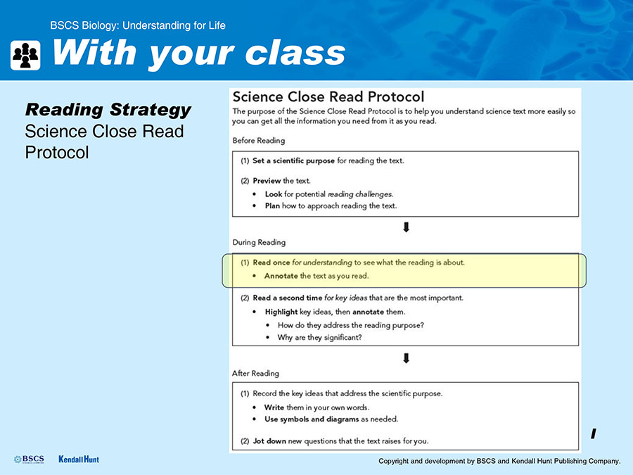
Describe and model the first read of the “during” reading phase of the close-reading protocol using the first five paragraphs of the reading.
Display Slide I. Explain that it takes a lot of different mental moves to read a science text for understanding. After you walk through this together, students will have the list of steps in their science notebooks to refer to in the future.
Read the first two paragraphs of the text, and share your thinking with think-alouds and annotations. As you annotate, use some of the Science Reading Annotation Stems.
Pause after reading the first portion of the text and ask students to describe what they noticed. Suggested prompt:
I just modeled what I do while I read. What did you notice about what I was doing? What could we call the strategies I was using?
Direct students to locate the Science Reading Annotation Stems handout in their science notebooks. Point out the annotation stems that you used and draw their attention to how they can use these kinds of thinking and writing stems as they annotate.
Invite students to read and annotate the next paragraph of text and accompanying graphic with their own reading process.
Ask students to pause after reading and invite students to share what they noticed and name the strategies they used to read the text, or some annotations they included.
Tell students to continue their first read of the text through the first paragraph of “The bad” subsection of the text, and then stop. Remind students to continue to refer to the Science Reading Annotation Stems list if they get stuck and need help.
During their independent reading time, pause students every so often and ask them to share their annotations and help each other make sense of the text.
Describe and model the second read of the “during” reading phase of the close-reading protocol using the first five paragraphs of the reading.
Display Slide J. Explain that in science reading, once we understand the meaning of a text, we need to consider how it supports our investigation by looking for key ideas to explain the difference between bacteria being on our bodies or in our bodies and our bodies being infected and answer the questions we came up with earlier (e.g., What environment(s) are the bacteria found in and how did they get there? What is the bacteria’s food source? How do the bacteria interact with other cells?)
Model identifying a piece of information in the text that may be a key idea. Ask students to think back to the lesson question and consider if there was anything here that helps with the questions we were trying to figure out.
Ask students to consider and name any strategies used to determine key ideas. Have them share their ideas with the class.
Ask students to reread the piece of text they already read once, and highlight and annotate key ideas. Once students are finished reading, invite students to share key ideas they found and explain why they think they are key ideas.
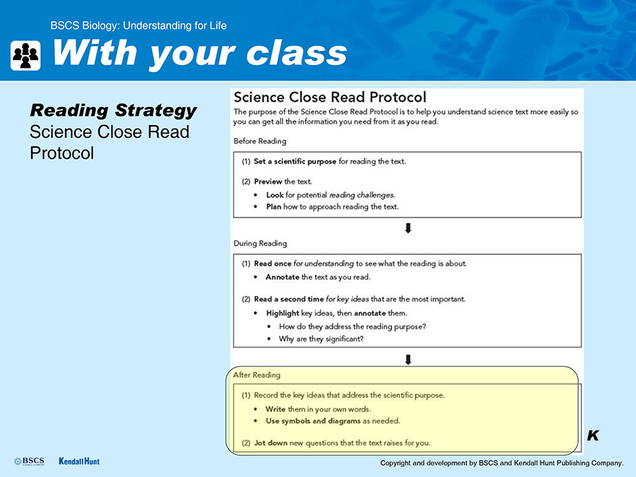
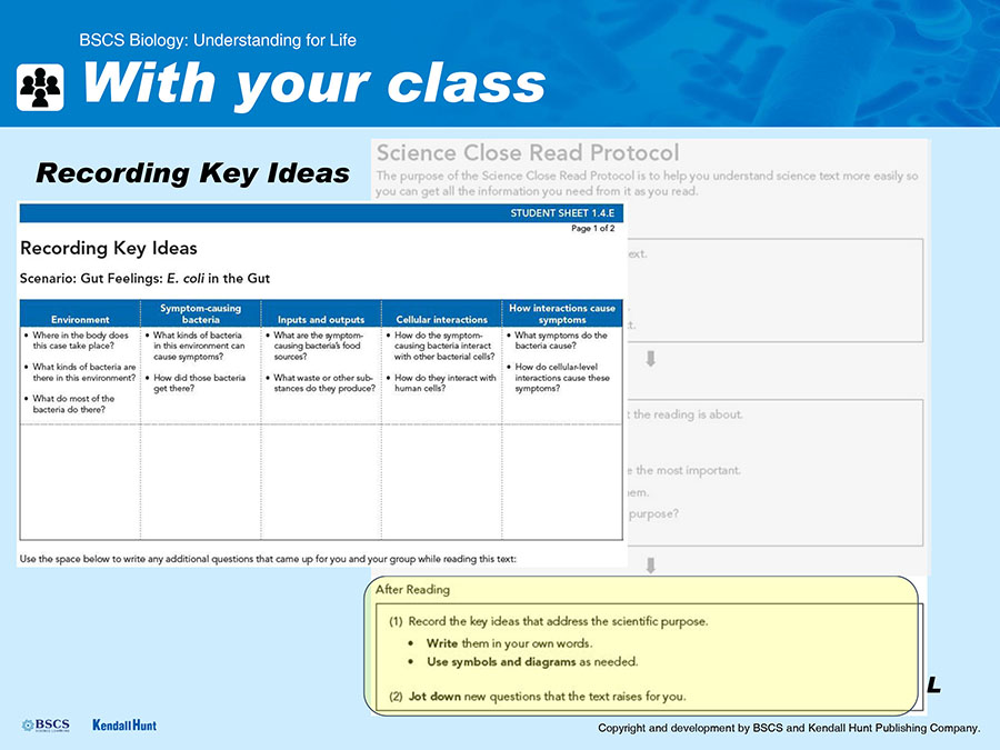
Explain and model the “after” reading phase of the close-reading protocol using the first five paragraphs of the reading.
Display Slide K. Distribute Student Sheet 1.4.E: Recording Key Ideas, which serves as a graphic organizer for the key ideas they identify. Display Slide L. Have students use the first side of the Recording Key Ideas graphic organizer to record key ideas from the first five paragraphs of the text. They should record information in their own words with symbols and diagrams as needed for each question they can answer in the “Environment” and “Symptom-causing bacteria” columns, and then record any questions that the text is raising for them. Depending on your students, you might choose to model your use of the graphic organizer to summarize key ideas for one of the questions.
 X
X
|
 X
X
|
Student groups are assigned scenarios.
Assign students to small groups (three or four students per group) and assign each group a scenario (ideally, you would have two groups looking at each scenario). Hand out a copy of Student Sheet 1.4.B: Bacteria in the Mouth, Student Sheet 1.4.C: Myth Busted, or Student Sheet 1.4.D: What Really Causes Acne? to each student, depending on group assignment. Students working on Student Sheet 1.4.A: Gut Feelings will continue and complete the reading from where you left off as a class using the text they have already started. You might choose to give specific scenarios to specific groups of students based on where you anticipate students might be challenged within the texts.
Common Student Ideas
Students may think that only certain tissues, like blood or brain tissue, are made of cells. These readings will expose students to a variety of body systems and tissues that are made up of cells.
Literacy and Multilingual Learner Support
Smaller group structures can be especially helpful for emerging multilingual learners because they give students a chance to engage in rich sensemaking with their peers and offer them the space to use their linguistic and nonlinguistic resources to express their ideas (and learn from other students’ uses of these resources, too).
Students use the science close-reading protocol to read through their assigned scenario with their groups.
Display Slide M. Remind students that they will be joining jigsaw groups during the next class, so they will need to become experts on their scenario in order to report about it to other students who will not have read it.
Remind students how the Science Close Read Protocol can be useful and support students to use it with their groups.
Remind students to use the Recording Key Ideas graphic organizer to remind them of the purpose of their reading as they preview the text and to record the key ideas in their own words and jot down any new questions the text raised.
Tell students they will report their responses from their Recording Key Ideas graphic organizer to their jigsaw group once everyone has finished using the science close read protocol to examine their scenario.
Keep Slide M displayed on the projector as students do this work to remind them of the information they will need to report back to their jigsaw group.
XAs students are working together in their groups, support and probe student thinking as well as their use of the close-reading protocol. Suggested prompts:
What did you notice about the text? Images? Graphics?
How are you refocusing your minds when they begin to wander?
What did you learn from the other members of your group by reading this together that you couldn’t have learned on your own?
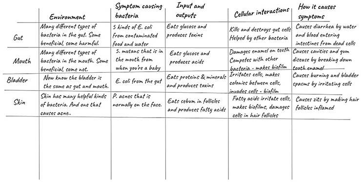
Sample of what students might list on their Recording Key Ideas graphic organizer for each of the four scenarios:
Scenario 1: Gut Feelings: E. coli in the Gut
Environment:
There are thousands of different bacteria in the gut; some of them are good, some are bad.
E. coli can be found in over 90% of human intestinal tracts.
Most bacteria in the gut grow as part of a healthy intestinal tract.
Symptom-causing bacteria:
A few strains of E. coli are harmful to humans; five strains seem to cause some form of diarrhea.
ETEC, EPEC, EIEC, EHEC, EAEC
They are mostly ingested from contaminated food or drink.
They can come directly from feces into the mouth.
Inputs and outputs:
Almost all strains of E. coli consume glucose (a simple sugar) present in the gut to fuel growth and reproduction.
E. coli produces toxins.
Cellular interactions:
Pathogenic E. coli organisms interact with nonpathogenic bacteria in the human gut that can help them cause more symptoms.
Some of the substances bacteria produce are toxic to human cells and destroy them, like Shiga toxin.
E. coli pokes holes in intestinal cells.
E. coli attaches to intestinal cells.
E. coli tells intestinal cells to build pedestals.
E. coli kills intestinal cells.
How interactions cause symptoms:
E. coli use of intestinal cells to grow and reproduce causes the cells to release water into the intestines, which eventually leads to diarrhea.
When E. coli kills cells, it sometimes causes bloody sores that can add blood to the diarrhea.
Sometimes the presence of other bacteria might make the E. coli cause more serious symptoms.
Scenario 2: Bacteria in the Mouth: The True Story of Why You Get Cavities
Environment:
There are hundreds of kinds of bacteria that naturally occur in our mouths.
Three kinds of Streptococcus bacteria are common in our mouths: S. gordonii, S. oralis, and S. mutans.
Most mouth bacteria don’t cause any harm.
Sometimes they form biofilms.
Symptom-causing bacteria:
A few of them become harmful in the right conditions.
Streptococcus mutans is the bacteria that causes cavities.
The bacteria live in the mouth from the time you’re a baby, but they don’t always cause problems.
Inputs and outputs:
S. mutans consume carbohydrates left on our teeth after we eat.
S. mutans produce acid as a waste product.
S. mutans thrive in a sugary acidic environment.
Cellular interactions:
S. mutans produce acid as waste. That acid causes our tooth enamel to deteriorate.
S. mutans form communities called biofilms or plaques that are really hard to brush off your teeth.
How interactions cause symptoms:
When S. mutans bacteria stick to teeth they can form pits that allow more bacteria to grow and produce acid.
If enough enamel deteriorates, we get cavities and gum disease.
Scenario 3: Myth Busted: E. coli in the Urinary Tract
Environment:
The urinary tract is not sterile!
Over 100 different species of bacteria have been found in the urinary tract.
Most live and grow there and are not harmful or are even beneficial, like L. iners.
Symptom-causing bacteria:
A few of the species can become harmful.
E. coli in the bladder can cause symptoms.
E. coli comes from the rectum, usually, by not cleaning well after going to the bathroom.
Inputs and outputs:
E. coli consumes proteins and minerals found in the bladder and the urine.
E. coli produces toxins.
Cellular interactions:
E. coli in the urinary tract forms communities called biofilms that help the bacteria stick to each other and the bladder walls, which makes them really hard to get rid of.
Pathogenic E. coli attach themselves to bladder wall cells and eventually move into the cell and reproduce.
They form colonies stuck together with proteins and sugar and then the bacteria invade neighboring cells.
How interactions cause symptoms:
The bacteria and the toxins irritate the cells.
The irritated cells cause spasms and a burning sensation when you urinate.
Scenario 4: What Really Causes Acne? No, It’s Not Eating Chocolate
Environment:
Thousands of bacteria occur naturally on our skin.
Most of the bacteria on our skin isn’t harmful. A few are harmful in the “right” environment.
Symptom-causing bacteria:
Many kinds of skin bacteria can cause problems in the “right” environment.
Propionibacterium acnes is the bacteria that causes acne.
P. acnes lives normally on most people’s faces, but it doesn’t always cause symptoms.
Inputs and outputs:
P. acnes consumes sebum and turns it into fatty acids that they release.
P. acnes grows and reproduces well in air-free, sebum-filled environments like blocked pores.
Cellular interactions:
P. acnes produces fatty acid as waste. These fatty acids irritate skin cells.
P. acnes forms communities called biofilms that help the bacteria stick together and to our skin, making them really hard to wash off.
P. acnes grows down inside hair follicles and damages the hair follicles.
How interactions cause symptoms:
When hair follicles become blocked with dead cells and sebum and then there is plenty of sebum for P. acnes to eat, they grow and reproduce and make fatty acids that irritate the cells and make the follicle inflamed. This causes pimples.
Developing the Practices: Obtaining, Evaluating, and Communicating Information
Use of the Science Close Read Protocol supports students to integrate information about how life processes of bacteria (e.g., what they do to grow and reproduce in their environment) can lead to symptoms during an infection in humans. The protocol also supports them in summarizing information as they write their ideas using the Recording Key Ideas graphic organizer after reading.
Support students to make sense of each other’s ideas and add words to the “words we encounter” section of the Word Wall.
As students read and discuss the information in their scenarios, terminology is likely to come up that is new or unfamiliar. As this happens, support students to make sense of new vocabulary. Add new vocabulary to the class “words we encounter” word list.
Literacy and Multilingual Learner Support
The “words we encounter” word list is provided not as a list of words to be pretaught but as words students may learn/begin to learn by encountering them in the text or resource. As students present their information from their scenario they can nominate words from their text to be placed on the “words we encounter” part of the Word Wall. Examples of “words we encounter” from each scenario are listed below. The words your students list may vary from those below based on what they think is new and important.
Gut Feelings: E. coli in the Gut
Microbiota
Aerobic
Pathogenic
Nonpathogenic
Enterotoxigenic (ETEC)
Enteropathogenic (EPEC)
Enteroinvasive (EIEC)
Enterohemorrhagic (EHEC)
Enteroaggregative (EAEC)
Bacteria in the Mouth: The True Story of Why You Get Cavities
Biofilm
Glucose
pH
Caries
Myth Busted: E. coli in the Urinary Tract
Sterile
Microbiota
Pathogenic
Uropathogenic
Pili
Biofilm
What Really Causes Acne? No, It’s Not Eating Chocolate
Microbiota
Commensal
Mutualistic
Colonizing
Sebum
Students reflect on using the Science Close Read Protocol.
Once students are finished reading and annotating the text, display Slide N. With their groups, ask students to consider how the close read protocol helped them to understand the text and name the strategies they used while reading. After considering with their groups, ask students to share their ideas, thoughts, and strategies with the whole class.
Be sure students have attached the Student Sheet 1.4.F: Science Close Read Protocol to a page at the back of their science notebooks to keep as a reference for future lessons.
Meta Moment
Because this is the first time students are using the science close-reading protocol, it will be helpful to ask students to take some time after working with it to consider how previewing and reading this text multiple times supported them to read the text with purpose and understand key ideas.
SUGGESTED CLASS PERIOD BREAK
End of Day 1
 GENERATE AN EXPLANATION
PURPOSE | To build understanding from evidence
Students reconvene with their original scenario group.
Have students reconvene with their original group that worked on one of the scenarios during the previous class. Give them a few minutes to briefly share and review the key ideas specific to their scenario that they wrote on Student Sheet 1.4.E: Recording Key Ideas. Remind them that they will be the only one in their jigsaw group who will have read their scenario and they should be prepared to share their findings.
Students move to jigsaw groups to share their findings.
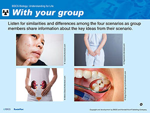
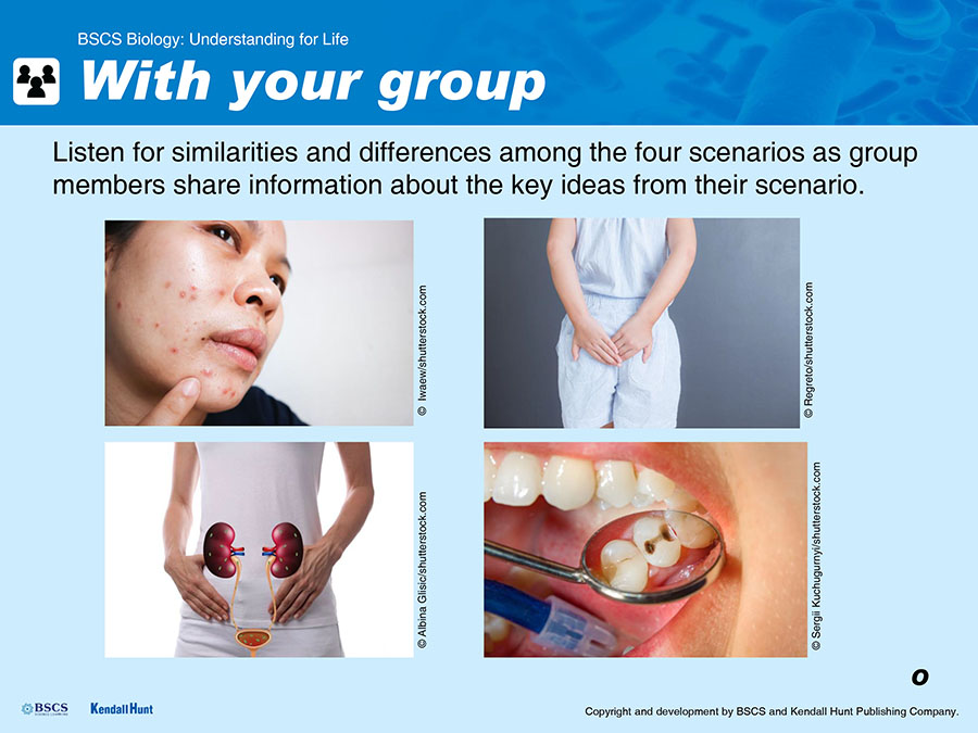
Display Slide O. Tell students we want to work together to figure out an answer to our lesson question about how bacteria cause us problems. Have students reassemble in groups of 4 in which each person has investigated a different one of the four scenarios. Students should take turns sharing what they learned in their individual scenarios and how the key ideas from their scenarios might help answer the lesson question about why some bacteria cause us problems.
Students set up a summary chart of their findings.
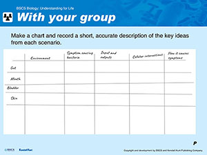
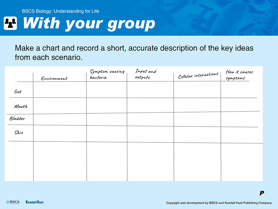
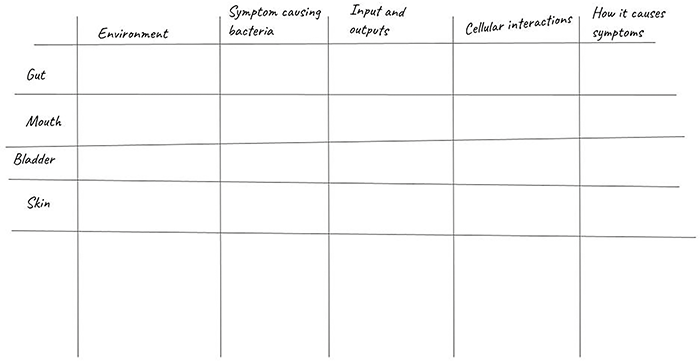
Display Slide P. Once everyone in the group has had a chance to share, distribute chart paper and markers to each group. Tell students to create a large chart on the chart paper that is similar to the one on Student Sheet 1.4.E: Recording Key Ideas with categories of questions across the top and space to label scenarios down the side. They should leave space at the bottom as shown to capture diagrammatic representations of their ideas after the next step.
Students paraphrase their findings.
For each of the scenarios, students in the group should work together to determine how to succinctly paraphrase the information from each scenario that answers the questions in each category, and record the paraphrased information in the appropriate places on the chart.
Environment: Where in the body does this case take place? What kinds of bacteria are there in this environment? What do most of the bacteria do there?
Symptom-causing bacteria: What kinds of bacteria in this environment can cause symptoms? How did those bacteria get there?
Inputs and outputs: What are the symptom-causing bacteria’s food sources? What waste or other substances do they produce?
Cellular interactions: How do the symptom-causing bacteria interact with other bacterial cells? How do they interact with human cells?
How interactions cause symptoms: What symptoms do the bacteria cause? How do cellular-level interactions cause these symptoms?
An example of what students may record is below.
Developing the Practices: Obtaining, Evaluating, and Communicating Information
Working together will support students as they work to summarize and paraphrase the scientific information that they obtained from the texts they read. Being able to paraphrase scientific information in simpler, but still accurate terms is an important element of communicating scientific information.
Developing the Crosscutting Concepts: Cause and Effect
See if students are suggesting that cause-and-effect relationships, such as life processes of bacteria (food consumption, production of waste and other substances, interaction with their environment and other cells) at a small scale, cause humans to experience symptoms of infection, at a larger scale.
Students reflect on developing scientific models.
Display Slide Q. Students’ goal is to come to a consensus about how to represent what we’ve figured out about each of the categories of key ideas, and record that diagrammatic representation in the space that is left at the bottom of their charts. Students spent some time learning about scientific models in Lesson 1, and have recorded their own models in the Model Tracker in Lessons 2 and 3. Remind them that scientific models are used to help explain how or why something works the way it does.
Developing the Practices: Developing and Using Models
The model elements that students are developing here should be thought of as initial or preliminary models. Students will have a chance to revise and reorganize these elements as they create their own models in their Model Trackers. The consensus model they’ll create as a class in Lesson 5 will give them another opportunity to refine and build on these ideas as they synthesize what they have learned in this chapter.
Students develop model representations.
Display Slide R.
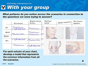
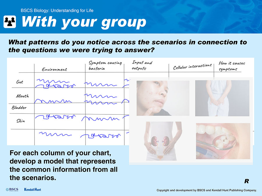
Students should work together to identify the common concepts or information in paraphrased summaries of the key ideas of all four scenarios. Then they should work together to decide how to represent that common information as they work toward developing a model that represents what they figured out in this lesson in a generalized way. Have them draw and label a diagram at the bottom of each column that shows how they think they might best represent:
Environment
Symptom-causing bacteria and where they come from
Inputs and outputs
Cellular interactions
How interactions cause symptoms
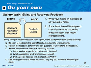
Prepare students for a gallery walk.
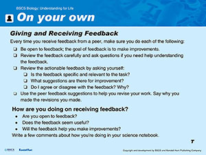
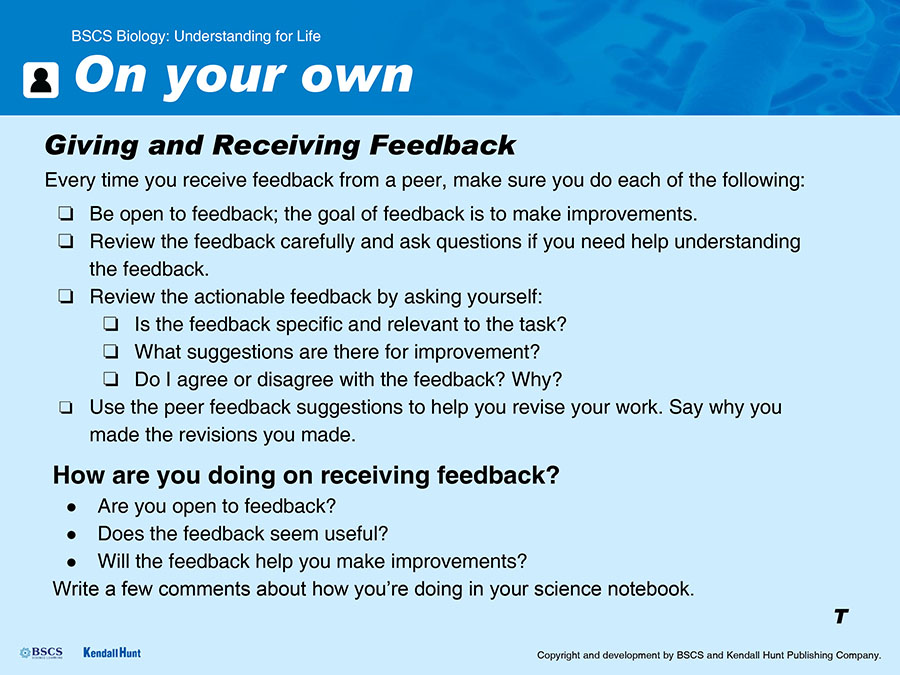
Display Slide S. In order to help students think about how they’ll create their own models in their Model Trackers, we will take a gallery walk to observe and give feedback on the representations that other groups have created. Distribute several sticky notes and a fine-tip marker to each student. Have them turn in their science notebooks to the page where they attached the Student Sheet 1.2.B: Giving and Receiving Feedback. Remind them that they will be respectfully providing specific and actionable feedback on the sticky notes similar to what we did in Lesson 2. Have them read through the receiving feedback portion of Student Sheet 1.2.B: Giving and Receiving Feedback. Remind them that they will be receiving feedback after the gallery walk. Allow students to ask any clarifying questions.
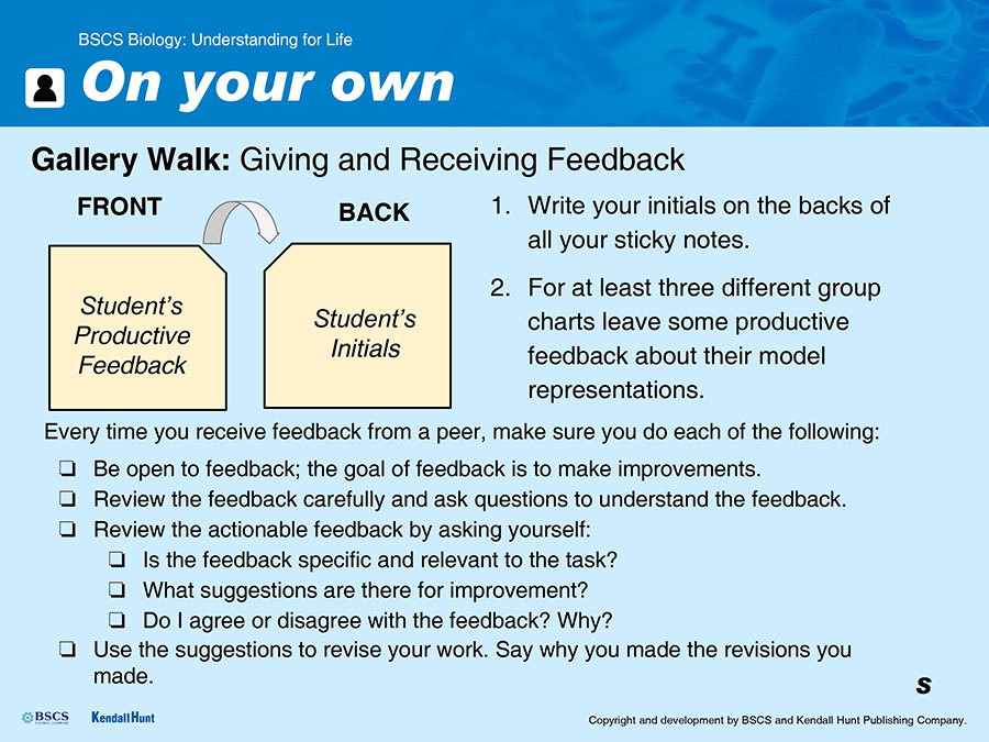
Students do a gallery walk and give feedback.
Display Slide S. Students should circulate among the charts made by other groups and observe the way they represented the key ideas. Everyone should give at least two feedback comments as they circulate among the charts made by other groups. Comments should be written on sticky notes, with their initials on the back, and then added to the appropriate charts as they circulate.
Students individually review feedback and receive feedback.
Display Slide T. Have students return to their jigsaw groups and direct them to silently read through the sticky notes that were attached to their chart. Ask them to recall the purpose of receiving feedback, referring to the handout as necessary, and to assess how they are doing individually at receiving feedback on the representations from their chart. Have them write a sentence or two about their reactions in their science notebooks.
Attending to Equity
Students may differ substantially in their ability and comfort level with receiving peer feedback and using it productively. Giving students a chance to reflect individually on their own experience and progress allows them to make adjustments and be prepared to engage with receiving peer feedback more publicly in future lessons.
Students discuss what we have figured out from our investigation.
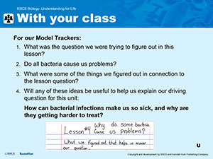
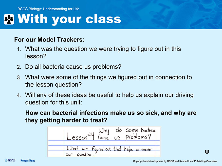
Display Slide U. Work through as a class what ideas we have figured out from our investigations during this lesson. Record what we figured out on a whiteboard or other publicly visible space.
| Suggested prompts | Listen for student responses such as |
|---|---|
| What was the question we were trying to figure out in this lesson? | Why do some bacteria cause us problems? |
| Do all bacteria cause us problems? |
|
| What were some of the things we figured out in connection to the lesson question? |
|
| Will any of these ideas be useful to help us explain our driving question for this unit: How can bacteria make us so sick? | Yes, all these ideas will help us understand how bacteria can make us so sick, especially the ones about how bacteria cause cell damage and impact body systems. |
If students bring up an idea that the class does not have complete consensus on, write down the idea and annotate it with a question mark to signal that we aren’t in complete agreement on this idea yet.
Students record the ideas we have figured out from the investigations in our Model Trackers.
Display Slide V. Prompt students to return to the Model Tracker in the front of their notebooks that they have been adding to in Lessons 2 and 3. Have them start an entry for Lesson 4 on the next fresh page, and record the lesson number, question, and the ideas we have figured out.
Have students think about the important ideas we figured out, and the feedback they got about how they represented these ideas on their charts, then work individually to draw a model in the last column that shows these ideas.
Potential ideas for representation can be seen in the image below:
Students use the Model Tracker Self-Assessment and Feedback Tool to evaluate and refine their representations.
Remind students to quietly consider the checklist criteria, review their models, and make any modifications they feel appropriate.
Formative Assessment Opportunity
Examine the Model Tracker entries for Lessons 2, 3, and 4 in student notebooks. The Model Tracker Formative Assessment Tool provides guidance on what to look for in student Model Trackers across all three of the lessons in the form of a checklist. As you work through the checklist keep in mind that you are looking for:
The presence or absence of components (not the quality of the representations)
Interactions that may be represented either in verbal descriptions or with diagrammatic elements such as arrows or lines
Interactions shown as arrows or other directional indicators showing inputs and outputs of bacterial cells (or other subsystems)
Interactions shown as arrows or otherwise indicating cause-and-effect relationships
Boundaries of subsystems in models from Lesson 4. These may not be explicitly indicated or labeled as system boundaries, but may be indicated simply by separating one subsystem (bacterial cell, human cell, tissue or organ that is affected) from another graphically or spatially. Subsystems should have indications of interactions between them.
Use of the model to explain something. Each lesson’s model will explain something a little bit different. The models should show some attempt to answer the lesson questions. For instance, a labeled bacterial cell alone is not sufficient in Lesson 2, rather the model should show some indication of how bacteria might grow.
Also keep in mind that you are NOT looking for neatness or for accuracy in the Model Tracker. The models students draw here are a description of their growing conceptual understanding and as such they may be inaccurate in some details of components, interactions, or explanations of phenomena.
In the appropriate space in each student’s Model Tracker Self-Assessment and Feedback Tool in the front of their science notebook, provide feedback to students regarding the models in their Model Tracker for Lessons 2–4. Give students feedback on:
Any missing, or extraneous components
The absence of any interactions that are described in the Assessment Tool
The directional nature of cause-and-effect relationships
Whether or not they have used the model to attempt to explain a phenomenon or answer a question (instead of simply labeling a diagram)
Any particularly effective elements of their models that helped you understand their thinking (Whether or not their thinking is accurate, the way they organize their models is as important as the accuracy of what they represent. This is why the feedback section has separate spaces for feedback on content and modeling.)
This feedback will help students improve their practice of modeling and support continued growth in their conceptual understanding.
Collect science notebooks from students for a formative assessment.
After class, review each student’s Model Tracker entries for this chapter using the Model Tracker Formative Assessment Tool available as an online resource. Enter individual feedback into that student’s Model Tracker Self-Assessment and Feedback Tool (attached at the very front page of the notebooks).
NAVIGATE
PURPOSE | To maintain coherence and continuity
Bring the class back together and facilitate a brief discussion about what we know so far that might be relevant to Zach’s case.
Suggested prompts:
Many of us are thinking that when bacteria can grow and reproduce in certain environments in our bodies they consume food sources, produce waste and other substances, and interact with human cells in ways that cause humans to feel symptoms of an infection.
But does this explain everything that happened to Zach?
How can we explain his case using the information we learned today?
Students add some words we’ve “earned” to the Word Wall.
Students have learned to distinguish between the harmless growth of nonpathogenic bacteria on us and in us and the growth of pathogenic bacteria that causes symptoms, which is an infection or an infectious disease. Add the following words to the Word Wall along with a visual representation of each word:
pathogenic (bacteria that cause symptoms)
nonpathogenic (bacteria that do not cause symptoms)
infection (the growth of pathogenic bacteria in a body)
infectious disease (disease caused by an infection)
Collect new questions that students have and add them to the DQB.
This discussion will be likely to raise a number of new questions for students about Zach’s case. Have students write any new questions they have from this discussion, or from earlier in the lesson, on sticky notes and add them to the DQB.
Ask students if they think it would be useful to pull together all the pieces of information we’ve collected in this chapter to help us explain Zach’s case.
Let them know that we will be putting the pieces together (all the ideas we figured out in this chapter) during the next class period.
REFERENCES
American Society for Microbiology. “E. coli’s Secret Weapon in Launching Infections.” ScienceDaily. August 20, 2019. www.sciencedaily.com/releases/2019/08/190820081857.htm (Student Sheet 1.4.A: Gut Feelings: E. coli in the Gut)
Evans, Doyle J. Jr., and Dolores G. Evans. “Escherichia coli in Diarrheal Disease.” In Medical Microbiology. 4th ed., edited by Samuel Baron. Galveston, TX: University of Texas Medical Branch at Galveston, 1996, chap. 25. https://www.ncbi.nlm.nih.gov/books/NBK7710 (Student Sheet 1.4.A: Gut Feelings: E. coli in the Gut)
Delmas, Julien, Guillaume Dalmasso, and Richard Bonnet. “Escherichia coli: The Good, the Bad and the Ugly.” Clinical Microbiology: Open Access 4, no. 2 (2015): 1–3. (Student Sheet 1.4.A: Gut Feelings: E. coli in the Gut)
Goswami, Kakolie, Chun Chen, Lingzi Xiaoli, Kathryn A. Eaton, and Edward G. Dudley. “Coculture of Escherichia coli O157: H7 with a Nonpathogenic E. coli Strain Increases Toxin Production and Virulence in a Germfree Mouse Model.” Infection and Immunity 83, no. 11 (2015): 4185–4193. (Student Sheet 1.4.A: Gut Feelings: E. coli in the Gut)
Hedlund, Kelly. “The Who’s Who of E. coli Strains.” Microbiologics. October 20, 2016. https://blog.microbiologics.com/the-whos-who-of-e-coli-strains (Student Sheet 1.4.A: Gut Feelings: E. coli in the Gut)
Taylor, Erika. “Why Are Some E. coli Strains Deadly While Others Live Peacefully in Our Bodies?” Scientific American. April 24, 2018. https://www.scientificamerican.com/article/why-are-some-e-coli-strains-deadly-while-others-live-peacefully-in-our-bodies (Student Sheet 1.4.A: Gut Feelings: E. coli in the Gut)
Friedman, Jason Yeshaya. “The Role of Streptococcus Mutans in the Formation of Dental Caries: An Ecological Perspective.” The Science Journal of the Lander College of Arts and Sciences 5, no. 1 (2011): 5. (Student Sheet 1.4.B: Bacteria in the Mouth)
Loesche, Walter J. “Role of Streptococcus Mutans in Human Dental Decay.” Microbiological Reviews 50, no.4 (1986): 353–380. (Student Sheet 1.4.B: Bacteria in the Mouth)
University of Chicago Dentistry. “The True Story of Why You Get Cavities, According to a Billion Microbes.” 2017. https://dentistry.uic.edu/patients/cavity-prevention-bacteria (Student Sheet 1.4.B: Bacteria in the Mouth)
Wanjek, Christopher. “The Truth About Tooth Decay.” LiveScience. November 6, 2007. https://www.livescience.com/2011-truth-tooth-decay.html (Student Sheet 1.4.B: Bacteria in the Mouth)
Anderson, Gregory G., Joseph J. Palermo, Joel D. Schilling, Robyn Roth, John Heuser, and Scott J. Hultgren. “Intracellular Bacterial Biofilm-Like Pods in Urinary Tract Infections.” Science 301, no. 5629 (2003): 105–107. (Student Sheet 1.4.C: Myth Busted: E. coli in the Urinary Tract)
Ackerman, Lenore A., and Toby C. Chai. “The Bladder is Not Sterile: An Update on the Urinary Microbiome.” Current Bladder Dysfunction Reports 14, no. 4 (2019): 331–341A. (Student Sheet 1.4.C: Myth Busted: E. coli in the Urinary Tract)
Gould, S.E. “Cystitis: How Bacteria Get Into Your Bladder.” Scientific American. October 7, 2012. https://blogs.scientificamerican.com/lab-rat/how-bacteria-invade-your-urinary-tract (Student Sheet 1.4.C: Myth Busted: E. coli in the Urinary Tract)
Hedlund, Kelly. “The Who’s Who of E. coli Strains.” Microbiologics. October 20, 2016. https://blog.microbiologics.com/the-whos-who-of-e-coli-strains (Student Sheet 1.4.C: Myth Busted: E. coli in the Urinary Tract)
Loyola University Health System. “New Findings on Bacteria in Female Bladders: Study Could Lead to Improved Diagnosis and Treatment of Urinary Tract Infections.” www.sciencedaily.com/releases/2018/06/180627160219.htm (Student Sheet 1.4.C: Myth Busted: E. coli in the Urinary Tract)
Thomas-White, Krystal, Samuel C. Forster, Nitin Kumar, Michelle Van Kuiken, Catherine Putonti, Mark D. Stares, . . . Trevor D. Lawley. “Culturing of Female Bladder Bacteria Reveals an Interconnected Urogenital Microbiota.” Nature Communications 9, no.1 (2018): 1–7. (Student Sheet 1.4.C: Myth Busted: E. coli in the Urinary Tract)
Wolfe, Alan J., Evelyn Toh, Noriko Shibata, Ruichen Rong, Kimberly Kenton, MaryPat FitzGerald, . . . Linda Brubaker. “Evidence of Uncultivated Bacteria in the Adult Female Bladder.” Journal of Clinical Microbiology 50, no. 4 (2012): 1376–1383. (Student Sheet 1.4.C: Myth Busted: E. coli in the Urinary Tract)
Macdonald, Fiona. “Researchers Might Have Figured Out Why Bacteria Only Causes Acne in Some People.” ScienceAlert. March 31, 2018. https://www.sciencealert.com/researchers-think-they-ve-finally-figured-out-why-bacteria-only-causes-acne-in-some-people
McLaughlin, Joseph, Steven Watterson, Alison M. Layton, Anthony J. Bjourson, Emma Barnard, and Andrew McDowell. “Propionibacterium Acnes and Acne Vulgaris: New Insights From the Integration of Population Genetic, Multi-omic, Biochemical and Host-Microbe Studies.” Microorganisms 7, no. 5 (2019): 128. (Student Sheet 1.4.D: What Really Causes Acne)
Sanford, James A., Ling-Juan Zhang, Michael R. Williams, Jon A. Gangoiti, Chun-Ming Huang, and Richard L. Gallo. “Inhibition of HDAC8 and HDAC9 by Microbial Short-Chain Fatty Acids Breaks Immune Tolerance of the Epidermis to TLR Ligands.” Science Immunology 1, no. 4 (2016): eaah4609. (Student Sheet 1.4.D: What Really Causes Acne)